
Implant Impression from a Laboratory Perspective
Implant impressions play a critical role in translating clinical conditions into a precise and functional restoration. From a dental laboratory perspective, the accuracy of the impression directly influences fit, contact, and overall restorative consistency. Factors such as proper seating of impression copings, stability during capture, and clarity of surrounding soft tissue all contribute to the final outcome. Both conventional and digital techniques are used, depending on case requirements and workflow.
Attention to detail during the impression stage can help reduce adjustments and remakes at the laboratory level. Elements such as adequate restorative space, accurate capture of implant position, and clear communication of case details support more predictable fabrication. While multiple clinical factors influence results, the quality of the impression remains a key step in the overall implant restoration process. Final clinical decisions should be based on professional judgment and individual case conditions.
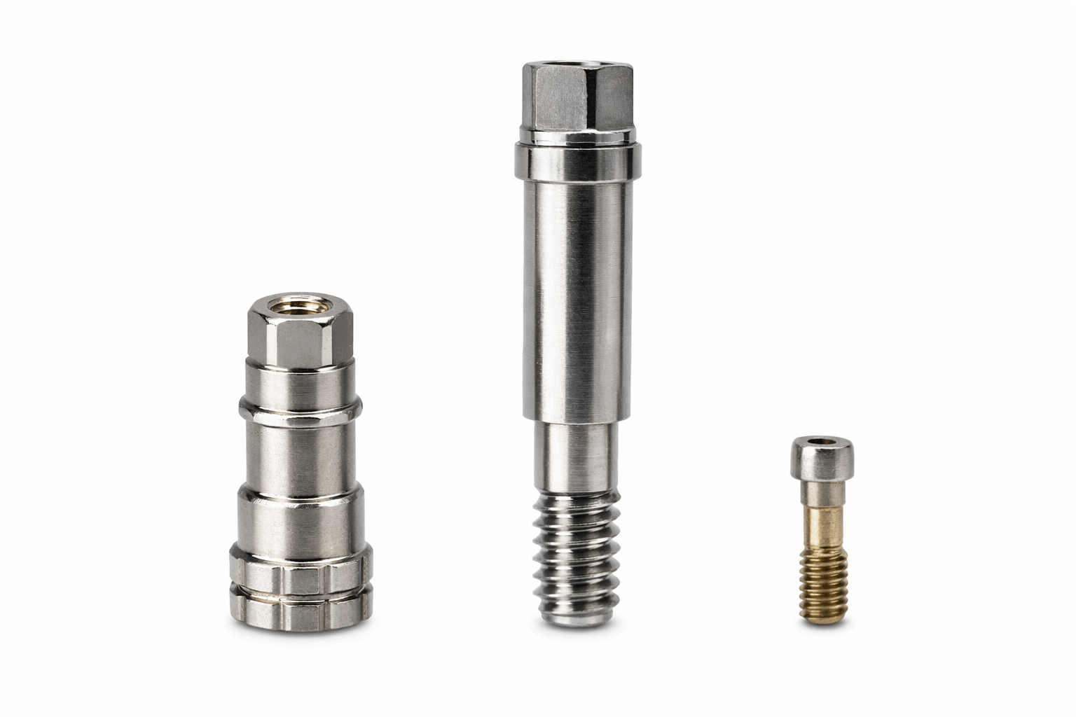
Types of Implant Impressions (Open Tray vs Closed Tray)
Implant impressions can be performed using open tray or closed tray techniques, depending on case requirements and clinical preference. Each method has specific indications and considerations. From a dental laboratory perspective, accurate transfer of implant position is critical to support consistent fit, alignment, and restorative outcomes.
Open Tray Impression Technique
Open tray impressions allow the impression coping to remain embedded within the impression when the tray is removed, providing a direct transfer of implant position. This technique is commonly used in multi-unit cases or when implants are angled, as it may reduce the risk of positional distortion. From a laboratory perspective, this can support more consistent model accuracy and alignment. However, the technique is more sensitive, requiring proper tray modification and access to the coping screw. It may also be less comfortable for some patients and can be more time-consuming. Accurate seating and stabilization during impression capture remain critical for predictable results.
Closed Tray Impression Technique
Closed tray impressions involve removing the impression tray while the coping remains in the mouth, followed by repositioning the coping into the impression. This method is often used for single-unit cases due to its simplicity and efficiency. It typically requires less chairside time and does not require tray modification. However, accuracy depends heavily on the correct repositioning of the coping into the impression. Any misalignment at this stage may affect the final model and restoration fit. From a laboratory perspective, clarity of the impression and precise transfer of implant position are essential to minimize discrepancies.
Selection Based on Case Conditions
The choice between open and closed tray techniques depends on clinical factors such as number of implants, angulation, access, and operator preference. Open tray impressions may be more suitable for complex or multi-unit cases where accurate transfer is critical, while closed tray impressions may be sufficient for straightforward single-unit restorations. From a dental laboratory perspective, consistency in technique and completeness of submitted information are more important than the method itself. Both approaches can produce reliable results when executed properly, but errors in seating, stability, or transfer may impact the final restoration outcome and require additional adjustments.
Digital Implant Impressions and Intraoral Scanning
Digital implant impressions use intraoral scanners to capture the implant position and surrounding structures without conventional materials. Scan bodies are used to transfer implant location into digital files for laboratory design. From a dental laboratory perspective, digital workflows may improve efficiency and communication, but accuracy depends on proper scanning technique, case conditions, and system compatibility.
Accuracy and Data Capture
Digital impressions can provide detailed visualization and eliminate some variables associated with conventional materials. Scan bodies allow the transfer of implant position into software for restoration design. In ideal conditions, digital capture may support consistent results, especially for single-unit cases. However, accuracy can be affected by scanning technique, soft tissue movement, and reflective surfaces. Incomplete data or stitching errors may impact final outcomes. From a laboratory perspective, clear and complete scans are essential, as missing or distorted data may require rescanning or adjustments during fabrication.
Efficiency and Workflow Integration
Digital workflows may improve efficiency by reducing material handling and allowing faster data transfer between the clinic and laboratory. Files can be reviewed immediately, and communication may be streamlined through digital platforms. This can support shorter turnaround times and easier case tracking. However, digital systems require proper training, equipment investment, and compatibility between scanner and laboratory software. Errors in scanning or file transfer may delay the process. From a laboratory perspective, consistent protocols and clear communication remain important to maintain workflow efficiency.
Case Selection and Limitations
Digital implant impressions may be well suited for single-unit cases with good access and stable soft tissue conditions. They can simplify the process and reduce patient discomfort in certain situations. However, multi-unit cases, deeply placed implants, or complex angulation may present challenges for digital capture. Limited access, saliva control, and soft tissue interference can affect scan accuracy. From a laboratory perspective, understanding when digital techniques are appropriate is important, as conventional impressions may still be preferred in certain complex cases to ensure reliable results.
Accuracy of Impression Copings and Components
The accuracy of impression copings and related components plays a critical role in transferring the implant position to the laboratory. Using the correct coping for the specific implant system, platform, and connection type is essential to support proper fit and alignment. From a dental laboratory perspective, mismatched or incorrectly selected components may lead to discrepancies in the working model and final restoration. Verification of part numbers, compatibility, and proper seating of the coping prior to impression capture can help reduce errors. Ensuring the coping is fully seated and secured, without movement during impression taking, is also important for accurate transfer. Incomplete seating or use of incorrect components may result in misfit, requiring adjustments or remakes. Careful selection and verification of all components before proceeding with the impression can contribute to more consistent restorative outcomes.
Importance of Proper Seating and Verification
Proper seating and verification are critical to accurately transfer implant position from the clinical setting to the laboratory. All protective covers or packaging components must be removed before impression procedures to avoid interference. Components should be fully seated and secured prior to capture. From a laboratory perspective, incomplete seating, incorrect assembly, or worn components can lead to discrepancies in the working model and final restoration. Careful verification at each step can reduce errors and improve overall consistency.
Removal of Protective Covers
All protective caps or packaging components must be removed before placing impression copings. These covers are designed for storage and protection, not for clinical use, and may prevent full seating if left in place. From a laboratory perspective, even minor interference at the interface can alter the recorded implant position. This may not be visible clinically but can result in discrepancies in the model and restoration. Verifying that all components are free of packaging materials prior to placement helps ensure accurate seating. A simple visual and tactile check before impression capture can prevent avoidable errors and support more predictable results.
Verification of Coping Seating
Impression copings must be fully seated and properly secured before taking an impression. Any gap, rotation, or instability can affect how the implant position is transferred to the laboratory. From a laboratory perspective, incomplete seating is one of the more common causes of misfit restorations. Clinicians may verify seating through radiographs, tactile confirmation, or manufacturer-specific guidelines. Ensuring that the coping is stable and correctly positioned prior to impression capture is essential. Consistent verification protocols can reduce the likelihood of discrepancies and minimize the need for adjustments or remakes during the restorative phase.
Use of Correct and Unworn Components
Using the correct components and avoiding reuse of impression copings, analogs, or related parts is important for maintaining accuracy. Even slight wear, which may not be visible, can affect how components fit together and transfer position. From a laboratory perspective, reused or worn components may introduce small discrepancies that become significant in the final restoration. Verifying part compatibility and using new, manufacturer-recommended components can help maintain consistency. Proper handling and selection of components support accurate model fabrication and reduce the risk of misfit in the final prosthesis.
Soft Tissue Management and Emergence Profile
Soft tissue management plays an important role in implant restoration, particularly in establishing a stable and functional emergence profile. From a dental laboratory perspective, the contour of the surrounding tissue influences how the restoration is designed and how it transitions from implant to crown. Proper use of healing abutments or provisional restorations may help shape soft tissue prior to final impression, supporting more natural contours and consistent margins. Inadequate tissue management can result in limited space, unclear margins, or compromised esthetics, which may affect restoration design. Accurate capture of soft tissue during impression or digital scanning is also critical to transfer the emergence profile correctly to the laboratory. While soft tissue outcomes depend on multiple clinical factors, early planning and management can support a more predictable restorative process and improve alignment between clinical conditions and laboratory fabrication.
Use of a Larger Healing Abutment for Tissue Shaping
Use of a larger healing abutment may assist in shaping the surrounding soft tissue to support a more favorable emergence profile prior to final impression. From a dental laboratory perspective, a well-developed tissue contour can allow for smoother transition from implant platform to restoration, improving both function and esthetic integration. Smaller healing components may result in tighter or less defined tissue, which can limit restorative design options. Selecting an appropriate size based on planned restoration can help guide tissue adaptation during healing. Adequate time should also be allowed for tissue stabilization before impression capture. While individual clinical conditions vary, thoughtful selection and use of healing abutments can contribute to more consistent soft tissue contours and support predictable laboratory fabrication of the final restoration.
Double-Cord Technique and Tissue Management
The double-cord technique is commonly used to manage soft tissue and improve margin visibility during impression procedures, and its principles remain relevant for both conventional and digital workflows. By placing two retraction cords, clinicians may achieve better lateral and vertical tissue displacement, allowing clearer access to margins or implant interfaces. From a dental laboratory perspective, improved tissue control can support more accurate capture of critical areas, whether using impression materials or intraoral scanning. Even in digital impressions, proper tissue management is important to avoid incomplete data or unclear boundaries. Inadequate retraction may result in distorted or missing information, affecting restoration design and fit. While technique and materials may vary, consistent tissue management supports a clearer definition of margins and emergence profile, contributing to more predictable outcomes in both conventional and digital implant restoration workflows.
Common Errors in Implant Impressions
Errors in implant impressions can affect the accuracy of the working model and the fit of the final restoration. From a dental laboratory perspective, common issues include incomplete seating of impression copings, movement during impression capture, and use of incorrect or incompatible components. Distortion of impression materials, inadequate soft tissue management, or unclear capture of implant interfaces may also contribute to discrepancies. In digital workflows, scanning errors such as missing data, stitching inaccuracies, or improper scan body positioning can affect results. Reuse of worn components or failure to verify proper assembly may introduce additional inaccuracies. These errors are often not immediately visible but can impact restoration fit, requiring adjustments or remakes. Careful attention to technique, component selection, and verification at each step can help reduce variability and support more consistent restorative outcomes.
Digital Implant Impressions and Intraoral Scanning
From Impression to Model: Laboratory Workflow
Digital implant impressions use intraoral scanners and scan bodies to capture implant position and surrounding structures without conventional materials. From a dental laboratory perspective, accurate scan body placement and complete data capture are essential for proper alignment and restoration design. Digital workflows may improve efficiency and communication, allowing immediate review and transfer of files. However, accuracy depends on scanning technique, access, soft tissue control, and system compatibility. Missing data, distortion, or improper scan body seating can affect outcomes. While digital methods may be suitable for many cases, proper case selection and technique remain important. Consistent protocols and verification of scan quality can support predictable results and reduce the need for rescanning or adjustments during fabrication.
After receiving an impression or digital file, the laboratory translates the captured data into a working model for restoration design. For conventional impressions, implant analogs are connected to impression copings and a model is fabricated to replicate intraoral conditions. For digital cases, scan data is processed and aligned to create a virtual model. From a dental laboratory perspective, accuracy at this stage depends on correct component use, proper seating, and completeness of the submitted information. Any discrepancies in the impression or scan may be transferred into the model and affect final fit. Careful verification and controlled workflow processes help support consistency in design, fabrication, and delivery of the final implant restoration.
Contact Us for Advanced Implant Planning
Early collaboration with the dental laboratory can support more consistent implant outcomes. We welcome dentists to contact us to discuss cases during the planning stage, particularly when evaluating restorative space, component selection, and workflow. From a laboratory perspective, early input may help align surgical placement with restorative requirements. Sharing digital images, intraoral scans, CBCT data, or case details can assist in preliminary evaluation and communication. This stage can be critical in identifying potential limitations and coordinating expectations before proceeding. While final clinical decisions remain with the treating provider, engaging the laboratory early may support a more predictable process from planning through final restoration.